Navigation auf uzh.ch
Navigation auf uzh.ch
Over the past decade, our group has strived to elucidate the molecular signals driving the puzzling concept of persistent cell death as a decisive factor of carcinogenesis. The liver and the intestine are both are characterized by a remarkable regenerative ability following tissue damage. This regenerative ability is dependent on a strict equilibrium between cell death and compensatory proliferation. Indeed, we have shown that interfering with this tightly regulated balance results in carcinoma development in both the liver and intestinal tract.
By genetically deleting the pro-survival protein MCL1 from either the liver or the intestine, we could demonstrate a crucial role for MCL1 in maintaining tissue homeostasis. Deletion of Mcl1 results in increased cell death, unregulated compensatory proliferation, DNA damage accumulation and carcinoma development. Human hepatocellular carcinoma (HCC) has several etiologies, leading to heterogeneous growth patterns and/or cytologic features within tumor that can ultimately challenge the tumor classification and thus compromise the treatment decision. By combining our pre-clinical models of HCC with patient derived samples, we are endeavoring to improve HCC characterization in respect to the contribution of hyperproliferation- and DNA damage-associated pathways.
The liver and intestinal tract are two of the most regenerative organs in the human body. As such, maintaining tissue homeostasis within these organs is reliant on a complex network of interacting cell death regulating molecules, disturbances of which are associated with cancer development. In recent years, the overexpression of MCL1, a potent anti-apoptotic protein of the BCL2 family, has been identified as a hallmark of many cancers, evidently enabling cancer cells to resist cell death. Interestingly however, we have shown that the deletion of MCL1 also results in impaired tissue homeostasis and eventual carcinoma development, indicating that MCL1 has both oncogenic and tumor suppressing functions within the liver and intestine.
This project aims at unraveling the modes by which MCL1 governs its homeostatic and tumor suppressing function in liver and gastro-intestinal tract. In particular, we strive to dissect what proportion of the pathology observed under MCL1 deficiency is generically due to pathologically increased apoptosis, and what specifically due to loss of MCL1 function.
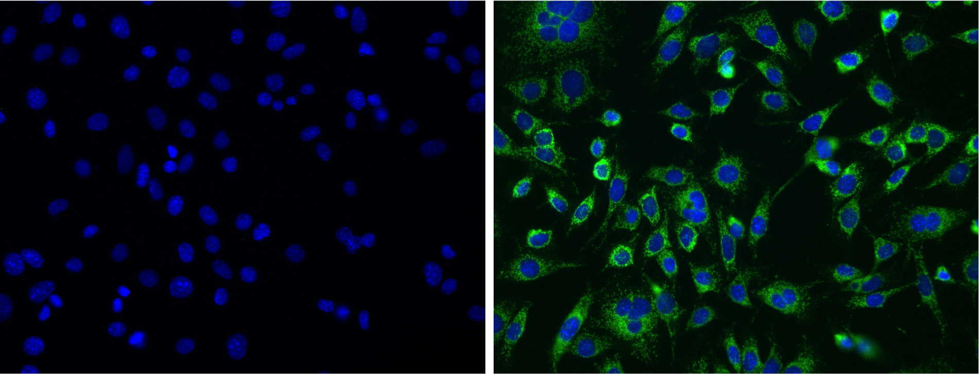
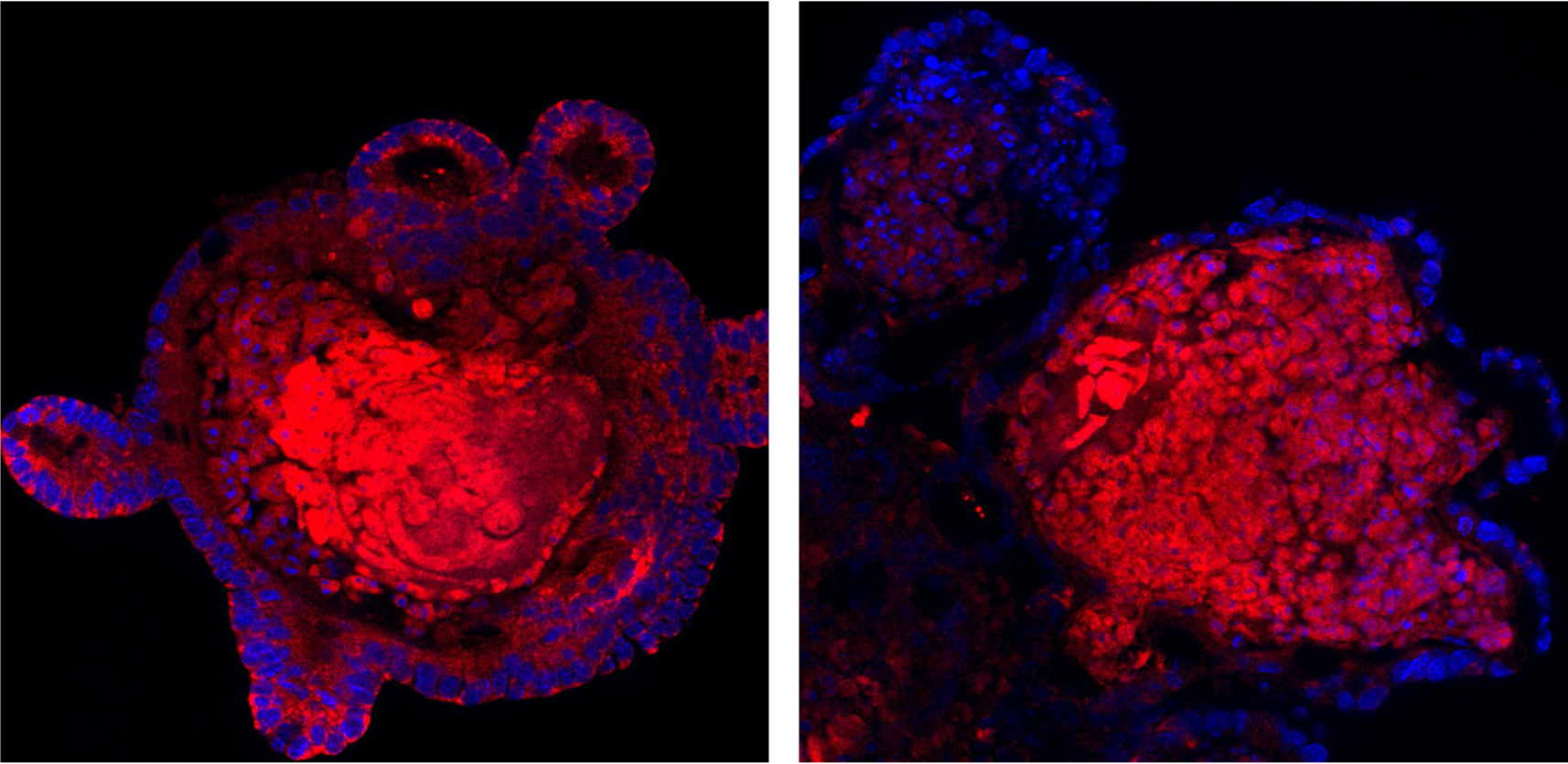
To this aim, we are engineering 2D and 3D cellular models that closely recapitulates the in vivo conditions of MCL1 deficiency in the liver and the gastro-intestinal tract enabling us to decipher the mechanism between lack of MCL1 and tumorigenesis.
Weber, A*, Boger, R*, Vick, B et al. (2010). Hepatocyte-specific deletion of the antiapoptotic protein myeloid cell leukemia-1 triggers proliferation and hepatocarcinogenesis in mice. Hepatology 51, 1226–1236.
Boege, Y, Malehmir, M, Healy, ME, et al. (2017). A Dual Role of Caspase-8 in Triggering and Sensing Proliferation-Associated DNA Damage, a Key Determinant of Liver Cancer Development. Cancer Cell 32, 342–359
Healy ME*, Boege Y*, Hodder MC*, et al. (2020). MCL1 Is Required for Maintenance of Intestinal Homeostasis and Prevention of Carcinogenesis in Mice. Gastroenterology 159(1):183-199.
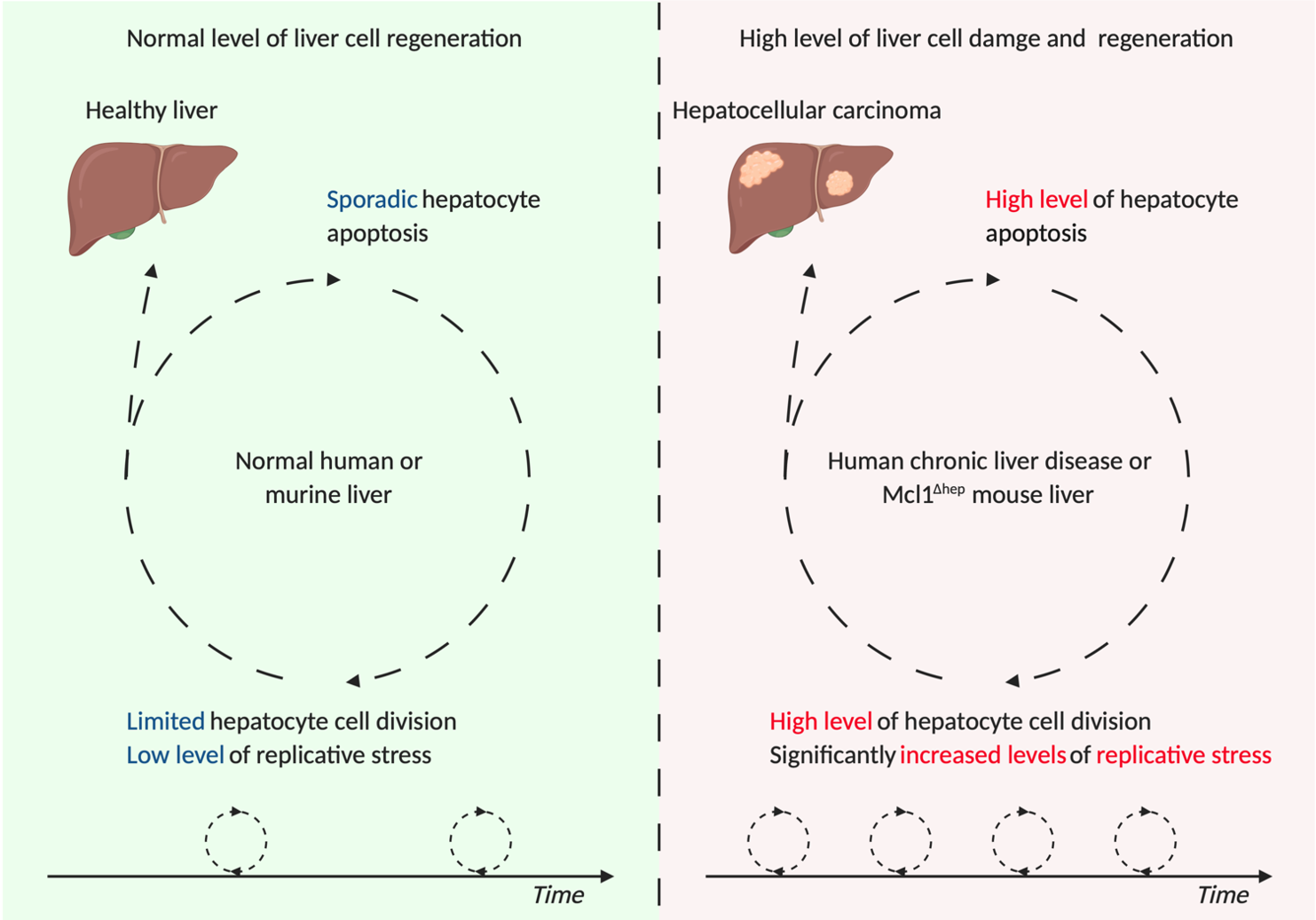
The liver is one of the few organs within the human body with complete regenerative capacity. This is not only essential for replacing hepatocytes which have been irreversibly damaged due disease or trauma but also facilitates the use of partial hepatectomy as one of our primary interventions in patients with liver cancer. In both cases, liver regeneration and restoration of function is driven by a strictly controlled increase in hepatocellular proliferation. However, increased hepatocellular proliferation requires a faster rate of cell division and an increased potential for replicative stress. Replicative stress is a major cause of genome instability, and is known to be a key driver of cellular transformation and the acquisition of tumorigenic properties.
In this project, conducted in close collaboration with the Lopes lab, we aim to investigate to what extent faster cell divisions under pro-apoptotic conditions, such as induced liver injury and/or MCL1 deficiency, lead to a higher probability of replication associated DNA damage and thus affect the integrity of the DNA of regenerating liver cells.
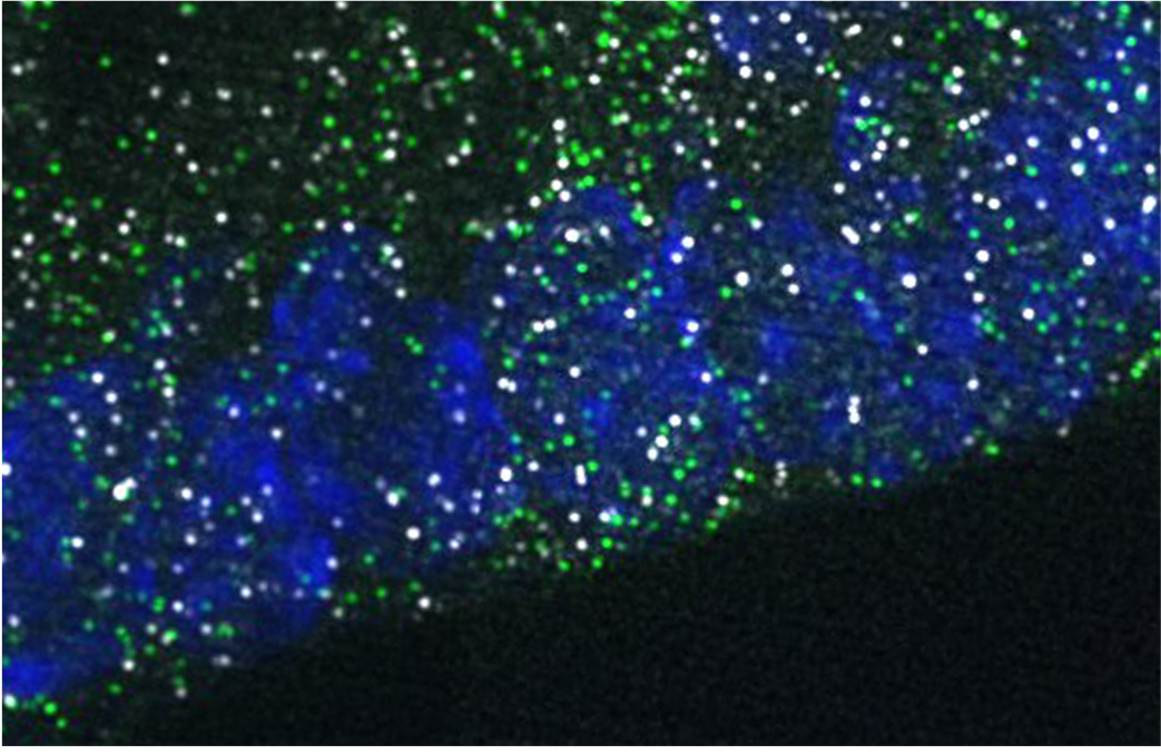
We are analyzing replication-associated DNA damage in liver tissues from several murine models of induced- acute and chronic liver damages, carrying or not MCL1 deficiency, by applying DNA fiber spreading analysis of individual fork progression. In addition, we apply electron microscopy to directly visualize potential replication fork remodeling.
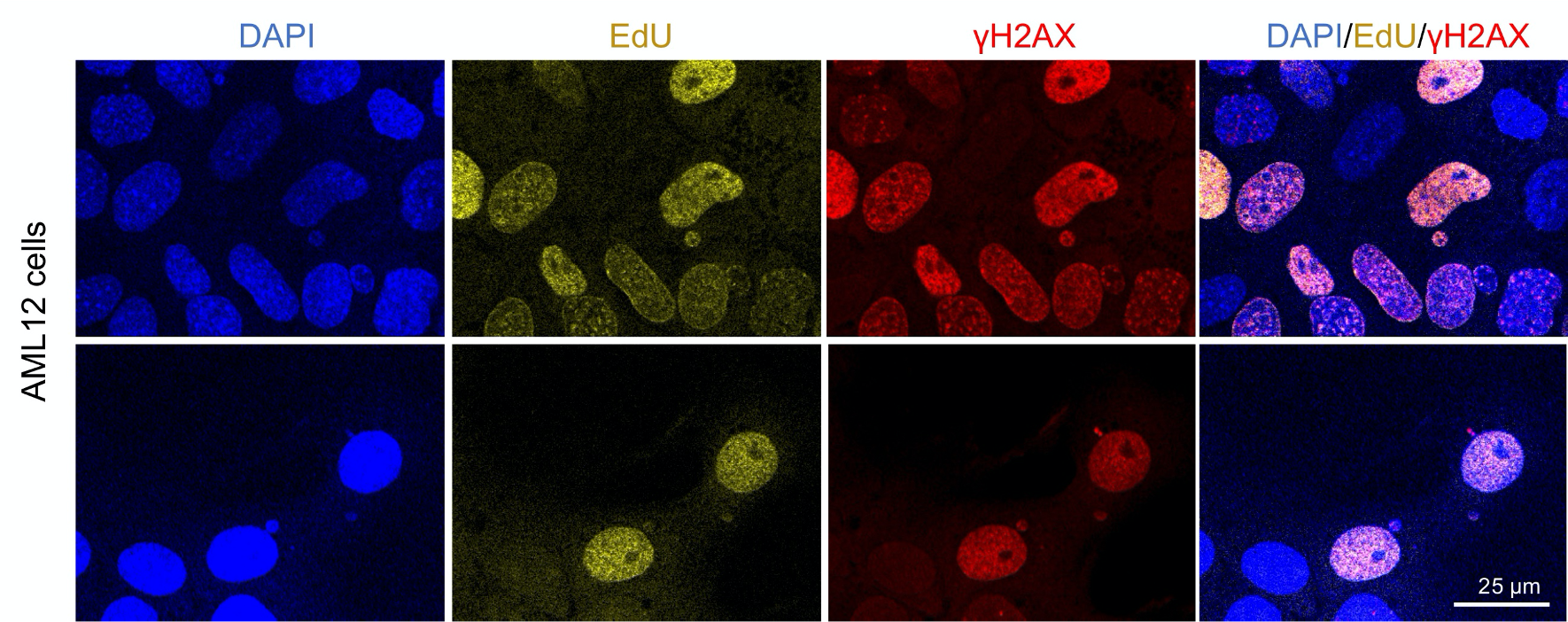
We are also assessing the impact of Mcl1 inactivation on the replication stress response by DNA fibers, electron microscopy and PFGE/comet assays in cell culture models upon genotoxic treatments.
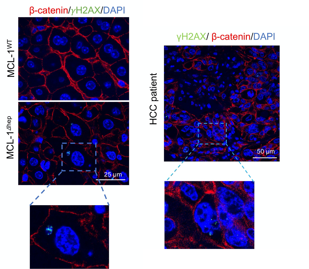
Regulated cell death, such as apoptosis, has been considered as a tumor suppressive mechanism to prevent an expansion of mutated cells. Evasion of such a mechanism is regarded as a hallmark of cancer. However, recent studies reveal a puzzling phenomenon that increased apoptosis can promote compensatory proliferation, consequential DNA replication stress and cancer development. We are interested in studying the mechanisms which bridge chronic liver injury and compensatory proliferation to liver tumorigenesis by using an animal model which lacks a key anti-apoptotic protein MCL1 in hepatocytes. MCL1 deficient livers display increased hepatocyte apoptosis which is followed by elevated compensatory proliferation, a phenomenon frequently observed in patients with chronic liver disease. At 12-months of age, 50% of MCL1 deficient mice develop hepatocellular carcinoma.
The aim of this study is to decipher the mechanisms on how chronic liver damage promotes compensatory proliferation in liver and how it modulates the mutational landscape. In parallel, we aim to determine the contribution of the cytosolic DNA-sensing pathway in bridging DNA damage to immune response and consequently the establishment of a chronic inflammatory microenvironment, which promotes ultimately liver tumorigenesis.
We are currently investigating the effect of blocking the cytosolic DNA sensing pathway in liver cancer development by using several genetically modified mouse lines. We also perform isolation of different fraction of cells from MCL1 deficient liver to investigate if there is a crosstalk between different cell types. In parallel, we also use hepatocyte cell lines and organoids to study the direct consequence of MCL1 deletion. We also verify the observations from our animal model on patient tissue to confirm if our study is of translational value.